Search

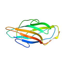
Solution structure of the FimH adhesin carbohydrate-binding domain
Authors
Van nuland, N.A., VANWETSWINKEL, S., Vranken, W.F., Buts, L.
Assembly
Solution structure of the FimH adhesin carbohydrate-binding domain
Entity
1. Solution structure of the FimH adhesin carbohydrate-binding domain (polymer, Thiol state: all disulfide bound), 158 monomers, 16907.74 Da Detail

FACKTANGTA IPIGGGSANV YVNLAPVVNV GQNLVVDLST QIFCHNDYPE TITDYVTLQR GSAYGGVLSN FSGTVKYSGS SYPFPTTSET PRVVYNSRTD KPWPVALYLT PVSSAGGVAI KAGSLIAVLI LRQTNNYNSD DFQFVWNIYA NNDVVVPT


Formula weight
16907.74 Da
Entity Connection
disulfide 1 Detail

IDTypeValue orderAtom ID 1Atom ID 2
1disulfidesing1:CYS3:SG1:CYS44:SG

Source organism
Escherichia coli
Exptl. method
solution NMR
Refine. method
RECOORD
Data set
assigned_chemical_shifts, heteronucl_T1_relaxation, heteronucl_T2_relaxation
Chem. Shift Complete
Sequence coverage: 100.0 %, Completeness: 97.3 %, Completeness (bb): 98.1 % Detail

Polymer type: polypeptide(L)

Total1H13C15N
All97.3 % (1710 of 1758)97.1 % (865 of 891)97.1 % (678 of 698)98.8 % (167 of 169)
Backbone98.1 % (910 of 928)98.1 % (313 of 319)97.6 % (450 of 461)99.3 % (147 of 148)
Sidechain96.9 % (945 of 975)96.5 % (552 of 572)97.6 % (373 of 382)95.2 % (20 of 21)
Aromatic92.9 % (156 of 168)92.9 % (78 of 84)92.7 % (76 of 82)100.0 % (2 of 2)
Methyl98.0 % (200 of 204)98.0 % (100 of 102)98.0 % (100 of 102)

1. FIMH

FACKTANGTA IPIGGGSANV YVNLAPVVNV GQNLVVDLST QIFCHNDYPE TITDYVTLQR GSAYGGVLSN FSGTVKYSGS SYPFPTTSET PRVVYNSRTD KPWPVALYLT PVSSAGGVAI KAGSLIAVLI LRQTNNYNSD DFQFVWNIYA NNDVVVPT

Sample

Solvent system 93% H2O/7% D2O, Pressure 1.000 atm, Temperature 298.000 K, pH 6.000, Details FimH_protonated 0.001000 M, sodium phosphate 0.020000 M, sodium chloride 0.100000 M


#NameIsotope labelingTypeConcentration
1FIMH[U-13C,15N]0.001 M
2sodium phosphatenatural abundance0.02 M
3sodium chloridenatural abundance0.1 M
4H2Onatural abundance93 %
5D2Onatural abundance7 %

LACS Plot; CA
Referencing offset: 0.04 ppm, Outliers: 3 Detail
LACS Plot; CB
Referencing offset: 0.04 ppm, Outliers: 3 Detail
LACS Plot; HA
Referencing offset: -0.12 ppm, Outliers: 2 Detail
LACS Plot; CO
Referencing offset: 0.69 ppm, Outliers: 2 Detail
Protein Blocks Logo
Calculated from 25 models in PDB: 3ZPD, Strand ID: A Detail


Heteronucl. T1
132 T1 values in 1 lists
Coherence Sz, Field strength (1H) 600 MHz, Pressure 1.000 atm, Temperature 298.000 K, pH 6.000 Detail
Heteronucl. T2
133 T2 values in 1 lists
Coherence S(+,-), Field strength (1H) 600 MHz, Pressure 1.000 atm, Temperature 298.000 K, pH 6.000 Detail
Heteronucl. T1/T2
132 T1/T2 values in 1 lists
Field strength (1H) 600 MHz, Pressure 1.000 atm, Temperature 298.000 K, pH 6.000 Detail
Release date
2014-02-09
Citation
Study of the structural and dynamic effects in the FimH adhesin upon α-d-heptyl mannose binding
Vanwetswinkel, S., Volkov, A.N., Sterckx, Y.G.J., Garcia-Pino, A., Buts, L., Vranken, W.F., Bouckaert, J., Roy, R., Wyns, L., van Nuland, N.A.J.
J. Med. Chem. (2014), 57, 1416-1427, PubMed 24476493 , DOI 10.1021/jm401666c ,
Related entities 1. Solution structure of the FimH adhesin carbohydrate-binding domain, : 1 : 63 : 3 : 6 : 26 entities Detail
Interaction partners 1. Solution structure of the FimH adhesin carbohydrate-binding domain, : 4 interactors Detail
Experiments performed 14 experiments Detail
nullKeywords bacterial adhesin, carbohydrate, CELL ADHESION, NMR, urinary tract infection