Search

Ribonuclease A in 40% acetic acid and 8M urea pH 2
Authors
L pez Alonso, J., Bruix, M., Laurents, D.
Assembly
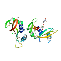
RNase A
Entity
1. RNase A (polymer, Thiol state: all disulfide bound), 124 monomers, 13690.17 Da Detail

KETAAAKFER QHMDSSTSAA SSSNYCNQMM KSRNLTKDRC KPVNTFVHES LADVQAVCSQ KNVACKNGQT NCYQSYSTMS ITDCRETGSS KYPNCAYKTT QANKHIIVAC EGNPYVPVHF DASV


Formula weight
13690.17 Da
Entity Connection
disulfide 4 Detail

IDTypeValue orderAtom ID 1Atom ID 2
1disulfidesing1:CYS26:SG1:CYS84:SG
2disulfidesing1:CYS40:SG1:CYS95:SG
3disulfidesing1:CYS58:SG1:CYS110:SG
4disulfidesing1:CYS65:SG1:CYS72:SG

Source organism
Bos taurus
Exptl. method
solution NMR
Data set
assigned_chemical_shifts
Chem. Shift Complete1
Sequence coverage: 100.0 %, Completeness: 55.6 %, Completeness (bb): 96.9 % Detail

Polymer type: polypeptide(L)

Total1H13C15N
All55.6 % (765 of 1376)38.5 % (275 of 714)70.9 % (372 of 525)86.1 % (118 of 137)
Backbone96.9 % (713 of 736)96.0 % (237 of 247)97.0 % (358 of 369)98.3 % (118 of 120)
Sidechain22.3 % (170 of 761) 8.1 % (38 of 467)47.7 % (132 of 277) 0.0 % (0 of 17)
Aromatic 3.2 % (3 of 94) 6.4 % (3 of 47) 0.0 % (0 of 47)
Methyl19.0 % (19 of 100) 8.0 % (4 of 50)30.0 % (15 of 50)

1. RNase A

KETAAAKFER QHMDSSTSAA SSSNYCNQMM KSRNLTKDRC KPVNTFVHES LADVQAVCSQ KNVACKNGQT NCYQSYSTMS ITDCRETGSS KYPNCAYKTT QANKHIIVAC EGNPYVPVHF DASV

Sample #1

Solvent system 40% acetic acid (deuterated), 50% H2O (milliQ), 10% D2O, Pressure 1 atm, Temperature 298 K, pH 2


#NameIsotope labelingTypeConcentration
1RNase A[U-13C]1.7 mM
2acetic acidnatural abundance40 %
3H2Onatural abundance50 %
4D2Onatural abundance10 %
Sample #2

Solvent system 8 M urea pH 2.5 10% D2O, Pressure 1 atm, Temperature 298 K, pH 2


#NameIsotope labelingTypeConcentration
5RNase A[U-100% 13C; U-100% 15N]1.7 mM
6D2Onatural abundance10 %
7ureanatural abundance8 M

Chem. Shift Complete2
Sequence coverage: 100.0 %, Completeness: 48.3 %, Completeness (bb): 83.0 % Detail

Polymer type: polypeptide(L)

Total1H13C15N
All48.3 % (1330 of 2752)31.6 % (451 of 1428)61.6 % (647 of 1050)84.7 % (232 of 274)
Backbone83.0 % (1222 of 1472)74.9 % (370 of 494)84.3 % (622 of 738)95.8 % (230 of 240)
Sidechain22.5 % (343 of 1522) 8.7 % (81 of 934)46.9 % (260 of 554) 5.9 % (2 of 34)
Aromatic 3.7 % (7 of 188) 5.3 % (5 of 94) 2.1 % (2 of 94)
Methyl21.5 % (43 of 200)12.0 % (12 of 100)31.0 % (31 of 100)

1. RNase A

KETAAAKFER QHMDSSTSAA SSSNYCNQMM KSRNLTKDRC KPVNTFVHES LADVQAVCSQ KNVACKNGQT NCYQSYSTMS ITDCRETGSS KYPNCAYKTT QANKHIIVAC EGNPYVPVHF DASV

Sample #1

Solvent system 40% acetic acid (deuterated), 50% H2O (milliQ), 10% D2O, Pressure 1 atm, Temperature 298 K, pH 2


#NameIsotope labelingTypeConcentration
1RNase A[U-13C]1.7 mM
2acetic acidnatural abundance40 %
3H2Onatural abundance50 %
4D2Onatural abundance10 %
Sample #2

Solvent system 8 M urea pH 2.5 10% D2O, Pressure 1 atm, Temperature 298 K, pH 2


#NameIsotope labelingTypeConcentration
5RNase A[U-100% 13C; U-100% 15N]1.7 mM
6D2Onatural abundance10 %
7ureanatural abundance8 M

LACS Plot; CA
Referencing offset: -0.21 ppm, Outliers: 5 Detail
LACS Plot; CB
Referencing offset: -0.21 ppm, Outliers: 5 Detail
Release date
2010-01-25
Citation
NMR spectroscopy reveals that RNase A is chiefly denatured in 40% acetic acid: implications for oligomer formation by 3D domain swapping
Lopez-Alonso, J., Bruix, M., Font, J., Ribo, M., Vilanova, M., Jimenez, M., Santoro, J., Gonzalez, C., Laurents, D.V.
J. Am. Chem. Soc. (2010), 132, 1621-1630, PubMed 20085318 , DOI 10.1021/ja9081638 ,
Entries sharing articles BMRB: 2 entries Detail
  BMRB: 16010 released on 2010-10-17
    Title How do Proteins Form Amyloid? Insight from the NMR Spectroscopic Characterization of 13C, 15N-labeled Ribonuclease A in 40% Acetic Acid
  BMRB: 16011 released on 2010-10-17
    Title NMR Spectroscopic Characterization of 13C, 15N-labeled Ribonuclease A in urea 8M
Related entities 1. RNase A, : 1 : 101 : 139 entities Detail
Interaction partners 1. RNase A, : 8 interactors Detail
Experiments performed 11 experiments Detail
Chemical shift validation 4 contents Detail
Keywords NMR Spectroscopy