Search

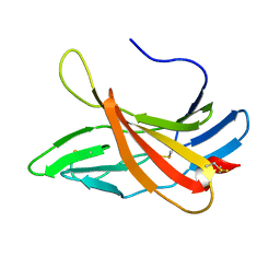
SOLUTION STRUCTURE OF CI-MPR domain 5 bound to N-acetylglucosaminyl 6-phosphomethylmannoside
Authors
Olson, L.J., Peterson, F.C., Volkman, B.F., Dahms, N.M.
Assembly
CI-MPR domain5
Entity
1. CI-MPR domain5 (polymer, Thiol state: all disulfide bound), 148 monomers, 16742.19 Da Detail

EAEAEFLSRT EGDNCTVFDS QAGFSFDLTP LTKKDAYKVE TDKYEFHINV CGPVSVGACP PDSGACQVSR SDRKSWNLGR SNAKLSYYDG MIQLTYRDGT PYNNEKRTPR ATLITFLCDR DAGVGFPEYQ EEDNSTYNFR WYTSYACP


Formula weight
16742.19 Da
Entity Connection
disulfide 3 Detail

IDTypeValue orderAtom ID 1Atom ID 2
1disulfidesing1:CYS15:SG1:CYS51:SG
2disulfidesing1:CYS59:SG1:CYS66:SG
3disulfidesing1:CYS118:SG1:CYS147:SG

Source organism
Bos taurus
Exptl. method
solution NMR
Refine. method
AUTOMATED METHODS WERE USED FOR BACKBONE CHEMICAL SHIFT ASSIGNMENT AND ITERATIVE NOE REFINEMENT. FINAL STRUCTURES WERE OBTAINED BY MOLECULAR DYNAMICS IN EXPLICIT SOLVENT
Data set
assigned_chemical_shifts
Chem. Shift Complete
Sequence coverage: 98.6 %, Completeness: 90.2 %, Completeness (bb): 95.8 % Detail

Polymer type: polypeptide(L)

Total1H13C15N
All90.2 % (1517 of 1682)90.6 % (790 of 872)87.7 % (575 of 656)98.7 % (152 of 154)
Backbone95.8 % (835 of 872)97.0 % (289 of 298)94.0 % (408 of 434)98.6 % (138 of 140)
Sidechain85.9 % (814 of 948)87.3 % (501 of 574)83.1 % (299 of 360)100.0 % (14 of 14)
Aromatic67.0 % (126 of 188)83.0 % (78 of 94)50.0 % (46 of 92)100.0 % (2 of 2)
Methyl96.6 % (112 of 116)96.6 % (56 of 58)96.6 % (56 of 58)

1. CI-MPR domain5

EAEAEFLSRT EGDNCTVFDS QAGFSFDLTP LTKKDAYKVE TDKYEFHINV CGPVSVGACP PDSGACQVSR SDRKSWNLGR SNAKLSYYDG MIQLTYRDGT PYNNEKRTPR ATLITFLCDR DAGVGFPEYQ EEDNSTYNFR WYTSYACP

Sample

Solvent system 95% H2O, 5% D2O, Pressure 1 atm, Temperature 308 K, pH 6.5, Details 1.25 mM domain-5 U-15N/13C, 8 mM N-acetylglucosaminyl 6-phosphomethylmannoside, 10 mM bis-Tris, 150 mM sodium chloride


#NameIsotope labelingTypeConcentration
1CI-MPR domain5[U-100% 13C; U-100% 15N]1.25 mM
2bis-tris[U-2H]10 mM
3sodium chloridenatural abundance150 mM
4N-acetylglucosaminyl 6-phosphomethylmannosidenatural abundance8 mM
5H2Onatural abundance95 %
6D2Onatural abundance5 %

LACS Plot; CA
Referencing offset: -0.42 ppm, Outliers: 1 Detail
LACS Plot; CB
Referencing offset: -0.42 ppm, Outliers: 1 Detail
LACS Plot; HA
Referencing offset: -0.14 ppm, Outliers: 1 Detail
LACS Plot; CO
Referencing offset: 0.48 ppm, Outliers: 2 Detail
Protein Blocks Logo
Calculated from 20 models in PDB: 2KVB, Strand ID: A Detail


Release date
2012-08-02
Citation
Structural basis for recognition of phosphodiester-containing lysosomal enzymes by the cation-independent mannose 6-phosphate receptor
Olson, L.J., Peterson, F.C., Castonguay, A., Bohnsack, R.N., Kudo, M., Gotschall, R.R., Canfield, W.M., Volkman, B.F., Dahms, N.M.
Proc. Natl. Acad. Sci. U. S. A. (2010), 107, 12493-12498, PubMed 20615935 , DOI 10.1073/pnas.1004232107 ,
Entries sharing articles BMRB: 1, Swiss-Prot: 1 entries Detail
  BMRB: 16772 released on 2012-08-02
    Title SOLUTION STRUCTURE OF CI-MPR ligand-free domain 5
  Swiss-Prot: P08169 released on 1988-08-01
    Title MPRI_BOVIN Entity Cation-independent mannose-6-phosphate receptor
Related entities 1. CI-MPR domain5, : 1 : 1 : 3 : 148 entities Detail
Interaction partners 1. CI-MPR domain5, : 39 interactors Detail
Experiments performed 3 experiments Detail
nullKeywords GLYCOPROTEIN, LYSOSOME, MANNOSE, MEMBRANE, NMR, PHOSPHOPROTEIN, RECEPTOR, SUGAR BINDING, TRANSMEMBRANE, TRANSPORT