Search

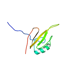
Structure of the UB2H domain of E.coli PBP1B in complex with LpoB
Authors
Simorre, J.P., Maya Martinez, R.C., Bougault, C.
Assembly
Penicillin-binding protein 1B (E.C.2.4.1.129,3.4.16.4)
Entity
1. Penicillin-binding protein 1B (E.C.2.4.1.129,3.4.16.4) (polymer, Thiol state: not present), 114 monomers, 13160.78 Da Detail

MGSSHHHHHH SSGLVPRGSH MGRMVNLEPD MTISKNEMVK LLEATQYRQV SKMTRPGEFT VQANSIEMIR RPFDFPDSKE GQVRARLTFD GDHLATIVNM ENNRQFGFFR LDPR


Formula weight
13160.78 Da
Source organism
Escherichia coli K-12
Exptl. method
solution NMR
Refine. method
simulated annealing
Data set
assigned_chemical_shifts
Chem. Shift Complete
Sequence coverage: 82.5 %, Completeness: 67.7 %, Completeness (bb): 72.3 % Detail

Polymer type: polypeptide(L)

Total1H13C15N
All67.7 % (908 of 1341)63.7 % (450 of 706)73.8 % (381 of 516)64.7 % (77 of 119)
Backbone72.3 % (486 of 672)69.6 % (160 of 230)74.6 % (249 of 334)71.3 % (77 of 108)
Sidechain65.4 % (507 of 775)60.9 % (290 of 476)75.3 % (217 of 288) 0.0 % (0 of 11)
Aromatic60.0 % (66 of 110)60.0 % (33 of 55)60.0 % (33 of 55)
Methyl87.0 % (80 of 92)89.1 % (41 of 46)84.8 % (39 of 46)

1. entity 1

MGSSHHHHHH SSGLVPRGSH MGRMVNLEPD MTISKNEMVK LLEATQYRQV SKMTRPGEFT VQANSIEMIR RPFDFPDSKE GQVRARLTFD GDHLATIVNM ENNRQFGFFR LDPR

Sample #1

Solvent system 90% H2O and 10% D2O, Pressure 1 atm, Temperature 293 K, pH 7.5, Details 400 uM [U-13C; U-15N] UB2H domain, 480 uM None LpoB, 90% H2O and 10% D2O


#NameIsotope labelingTypeConcentration
1UB2H domain[U-13C; U-15N]400 (±5.0) uM
2LpoBnatural abundance480 (±5.0) uM
3Tris.HClnatural abundance10 mM
4NaClnatural abundance200 mM
Sample #2

Solvent system 90% H2O/10% D2O, Pressure 1 atm, Temperature 293 K, pH 7.5, Details 450 uM [U-15N] UB2H, 540 uM None LpoB, 90% H2O/10% D2O


#NameIsotope labelingTypeConcentration
5LpoBnatural abundance540 uM
6UB2H[U-15N]450 (±5.0) uM
7Tris.HClnatural abundance10 mM
8NaClnatural abundance200 mM

LACS Plot; CA
Referencing offset: -0.19 ppm, Outliers: 2 Detail
LACS Plot; CB
Referencing offset: -0.19 ppm, Outliers: 2 Detail
LACS Plot; HA
Referencing offset: -0.06 ppm, Outliers: 3 Detail
LACS Plot; CO
Referencing offset: -0.05 ppm, Outliers: 2 Detail
Protein Blocks Logo
Calculated from 20 models in PDB: 6G5R, Strand ID: A Detail


Release date
2019-03-12
Citation 1
Structural Insights into Inhibition of Escherichia coli Penicillin-binding Protein 1B
King, D.T., Wasney, G.A., Nosella, M., Fong, A., Strynadka, N.C.
J. Biol. Chem. (2017), 292, 979-993, PubMed 27899450 , DOI 10.1074/jbc.M116.718403 ,
Citation 2
Induced conformational changes activate the peptidoglycan synthase PBP1B
Egan, A.J.F., Maya-Martinez, R., Ayala, I., Bougault, C.M., Banzhaf, M., Breukink, E., Vollmer, W., Simorre, J.P.
Mol. Microbiol. (2018), 110, 335-356, PubMed 30044025 , DOI 10.1111/mmi.14082 ,
Related entities 1. Penicillin-binding protein 1B (E.C.2.4.1.129,3.4.16.4), : 1 : 1 : 9 : 18 entities Detail
Interaction partners 1. Penicillin-binding protein 1B (E.C.2.4.1.129,3.4.16.4), : 10 interactors Detail
Experiments performed 12 experiments Detail
nullKeywords Complex, E. coli, Glycosyltransferase, LpoB, Murein polymerase, PBP1B, Peptidoglican, TRANSFERASE, Transpeptidase